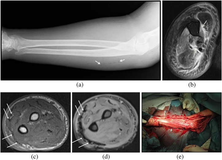
Figure 3.
Necrotizing fasciitis in the right upper limb of a 71-year-old male due to gas-forming organisms (Streptococcus anginosus). Radiograph shows faint air lucencies within the soft tissues (white arrows). (b) Axial T2 weighted MR image shows thick hyperintense signal in the deep intermuscular fascia (white arrows). (c) Axial T1 weighted MR image taken at a more distal level shows few tiny foci of signal void in the superficial fascia (white arrows). (d) Axial gradient-echo image shows blooming artefacts in the superficial fascia (white arrows) compatible with air foci—these are more apparent on the gradient-echo image than the T2 weighted image. (e) Urgent fasciotomy and surgical debridement was performed. Cultures grew Streptococcus anginosus.

