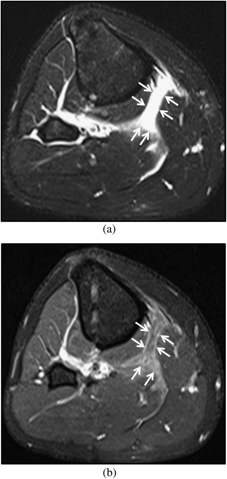
Figure 4.
Necrotizing fasciitis of the leg in a 21-year-old male with no underlying risk factors. (a) Axial fat-suppressed T2 weighted MR image of the mid-calf shows thick hyperintense fluid signal in the intermuscular fascia abutting the gastrocnemius muscle (white arrows). (b) Corresponding axial post-contrast fat-suppressed T1 weighted MR image showing mixed enhancing and non-enhancing areas, corresponding to T2 hyperintensity (white arrows). At surgery, early necrotizing fasciitis involving the fascia between superficial posterior and deep posterior compartments was found.

