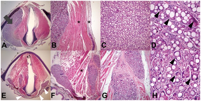
Figure 5. H&E staining of axial cross-sections of the larynx 12 months after injection with PCL microspheres (A–D) or CaHA (E–H).
Injected PCL microspheres remained at the left vocal fold (A, ×12.5, gray arrow). In contrast, the CaHA migrated from the left vocal fold, through the fascia space, and to the right vocal fold (E, ×12.5, white arrowheads). Injected PCL (B) and CaHA (F) microspheres did not cause an inflammatory reaction in the surrounding epithelium, lamina propria, or muscle (×40, asterisks). Giant cell formation occurred to the same extent in the PCL (D, ×400, black arrowheads) and CaHA groups (H, ×400, black arrowheads).

