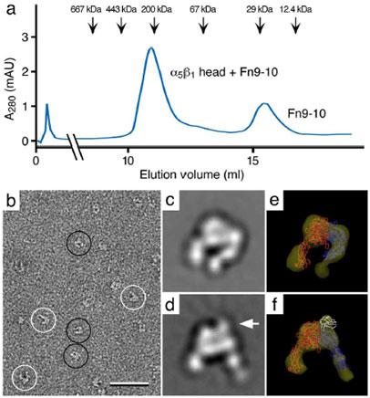
Fig. 3. Negative stain electron microscopy of the integrin α5β1 headpiece with and without a bound fibronectin (Fn) fragment containing Fn domains 7 to 10 (Fn9-10).

a: Elution profile from a gel filtration column used to purify the complex of α5β1 headpiece with an Fn9-10 fragment. The elution profile shows two peaks that correspond to the α5β1-Fn9-10 complex (~200 kDa) and unbound Fn9-10 fragment (~30 kDa). b: Negative stain electron microscopy reveals that the α5β1 headpiece adopts two conformations, namely a closed (black circles) and an open conformation (white circles). c and d: Class averages representing the closed (c) and the open conformation (d). Binding of Fn9-10 fragment (arrow in d) induces the open conformation of the headpiece, while the unliganded is in the closed conformation (c). e and f: 3D reconstructions of an unliganded (e) and an Fn9-10-liganded α5β1 headpiece (f) with the fit atomic structures of the αV and β3 subunit (33) in red and blue, respectively, and of the Fn9-10 fragment (34) in white. The scale bar corresponds to 50 nm and panels c to f have a side length of 22 nm. Figure panels modified and reprinted from (10). Copyright 2003 with permission from EMBO Journal.
