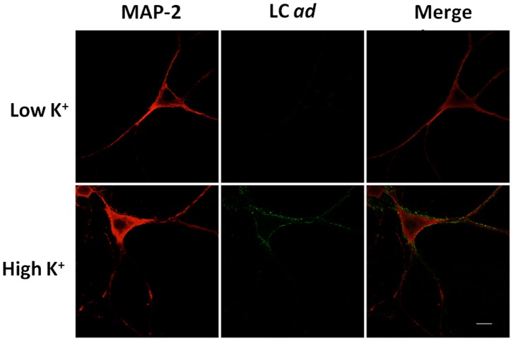
Figure 1. Immunofluorescence analysis showing BoNT/A ad binding during neuronal depolarization (active neurons).
E19 rat hippocampal neurons cultured in maintenance medium for 10/A ad for 1 min at 37°C in HEPES Ringer Resting Buffer (top row) or in high K+ HEPES Ringer Depolarization Buffer (bottom row). Anti-MAP-2 chicken monoclonal antibody (Cat # PCK-554P, Covance) was used to stain neuronal soma (red), and F1-40 antibody was used for detection of LC ad (green). Scale is 10 µm.

