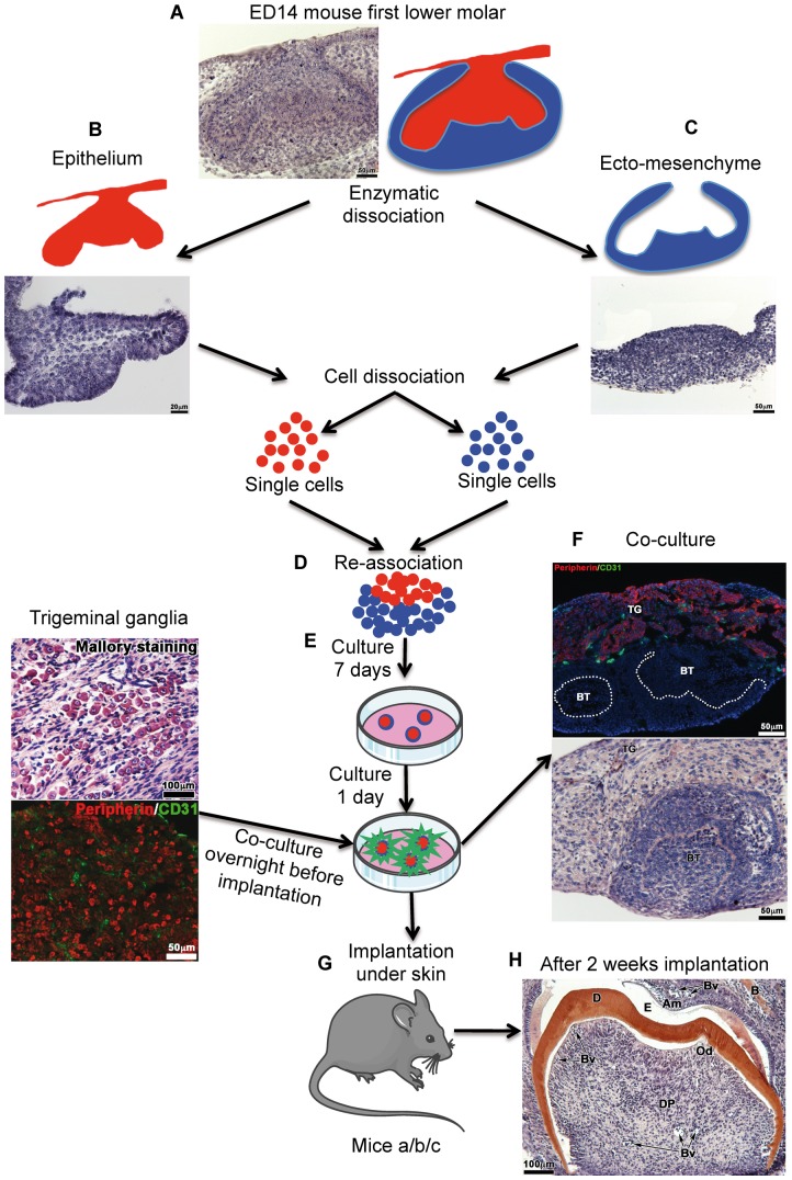
Figure 1. Protocol for tooth organ engineering.
The mandibular first molars were dissected from ICR mouse embryos at embryonic day (ED) 14 (cap stage) (A). Then, the dental epithelium (B) and ecto-mesenchyme (C) were separated by using a mixture of 0.25% trypsin and 1.2 U/mL dispase in DMEM-F12 (preheated to 37°C) at room temperature during 15 min. Each tissue was dissociated into single cells, which were then re-associated (D) and grown on semi-solid cultured medium (E). After 7 days in vitro, each re-association was co-cultured overnight with trigeminal ganglia from ICR newborn mice (F). The eighth day (G), bioengineered tooth unit and trigeminal ganglia were co-implanted between skin and muscles behind the ears in adult ICR mice (mice a), CsA-treated ICR mice (mice b) and Nude mice (mice c) for 1 week or 2 weeks (H). BT, bioengineered tooth; TG, trigeminal ganglia.

