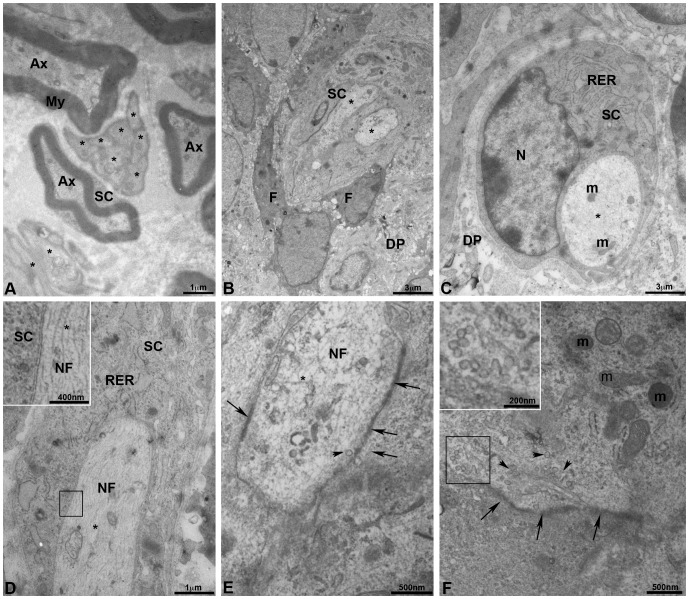
Figure 4. Innervation of bioengineered teeth implanted in cyclosporin A-treated ICR mice by transmission electron microscopy.
Transmission electron microscopy (TEM) of trigeminal ganglia (A) showed the presence of myelinated and unmyelinated axons surrounded by Schwann cells (A). TEM of dental pulp of epithelial and mesenchymal cell-cell re-associations co-implanted for 2 weeks with trigeminal ganglia in CsA-treated ICR mice showed the presence of unmyelinated axons (B). These axons were surrounded by a Schwann cell and located near fibroblasts, which secreted collagen (B). In C, one unmyelinated axon was surrounded by a Schwann cell with a developed rough endoplasmic reticulum. Neurofilaments (D, E), numerous secretory vesicles (arrowheads in F and insert) and mitochondria (F) were present in the axons. A typical structure of a pre-synapse with numerous mitochondria and synaptic vesicles (arrowheads) was observed (F). Thickening of the membrane suggested presence of synaptic contacts (arrows in E and F). Ax, myelinated axon; DP, dental pulp; F, fibroblasts; m, mitochondria; N, nucleus; NF, neurofilaments; My, myelin; RER, rough endoplasmic reticulum; SC, Schwann cells; *, unmyelinated axon.

