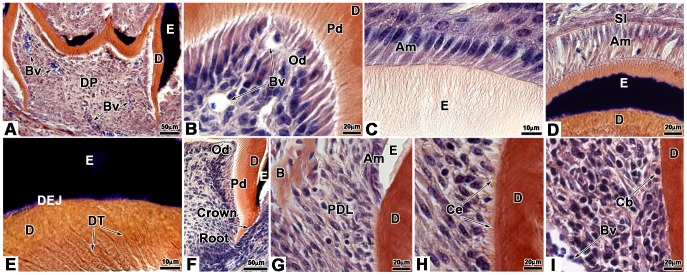
Figure 5. Histology of bioengineered teeth implanted for 2 weeks in cyclosporin A-treated ICR mice.
Bioengineered teeth implanted for 2 weeks under the skin in CsA-treated ICR mice were analysed by histology. Crown development was identical to that observed for Nude mice (A). Blood vessels could enter in the dental pulp (A), migrated in the pulp (A) and reached odontoblasts (B). Odontoblasts were elongated and polarized by the position of the nucleus, opposite to the secretory pole (B). Thus the secretion of predentin/dentin was polarized (B). As well as to Nude mice, ameloblasts of bioengineered teeth implanted in CsA-treated ICR mice, were elongated, polarized and secreted enamel (C). In contact with ameloblasts stratum intermedium could be observed (D). In the predentin/dentin, dentinal tubules were present and reached the dentin-enamel junction (E). After 2 weeks of implantation, root was developed (F) and newly formed bone was present in dental mesenchyme (G) as well as in Nude mice. In contact with the external surface of dentin, cementoblasts were observed (I). These cells were functional and deposited cementum (H). Periodontal ligament was attached to the root by cementum and extended until reaching newly formed bone (G, H). Am, ameloblast; B, bone; Bv, blood vessel; Cb, cementoblast; Ce, cementum; D, dentin; DEJ, dentin-enamel junction; DP, dental pulp; DT, dentinal tubule; E, enamel; Od, odontoblast; Pd, predentin; PDL, periodontal ligament; SI, stratum intermedium.

