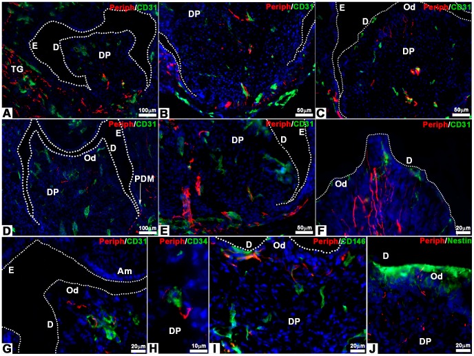
Figure 7. Innervation of bioengineered teeth implanted in Nude mice.
Bioengineered teeth germs were co-implanted with trigeminal ganglia in adult Nude mice (A–J) for 1 (A–C) or 2 weeks (D–J). Nerve fibers and blood vessels in dental pulp and peridental tissues of bioengineered teeth were analysed immunohistochemically by using specific antibodies for peripherin and CD31. Blood vessels were present in peridental tissues and could enter in the dental pulp and reach odontoblasts already after 1 week of implantation (A–C). The staining for peripherin showed that nerve fibers entered in the dental pulp (A, D) and extend in the pulp (B, E) after 1 (A–C) and 2 (D–J) weeks. After 1 week of implantation, nerve fibers did not reach the odontoblasts (C). This was achieved only after 2 weeks of implantation (F, G, I, J). Double staining for peripherin (G–I), CD31 (G), CD34 (H) and CD146 (I) showed associations between nerve fibers and blood vessels in the dental pulp (H) and subodontoblastic layer (G, I). After 2 weeks of implantation, nerve fibers, visualized by anti-peripherin antibody, had reached the odontoblast (positive for nestin) layer (J). Am, ameloblasts; D, dentin; DP, dental pulp; E, enamel; Od, odontoblasts; PDM, peridental mesenchyme; TG, trigeminal ganglia.

