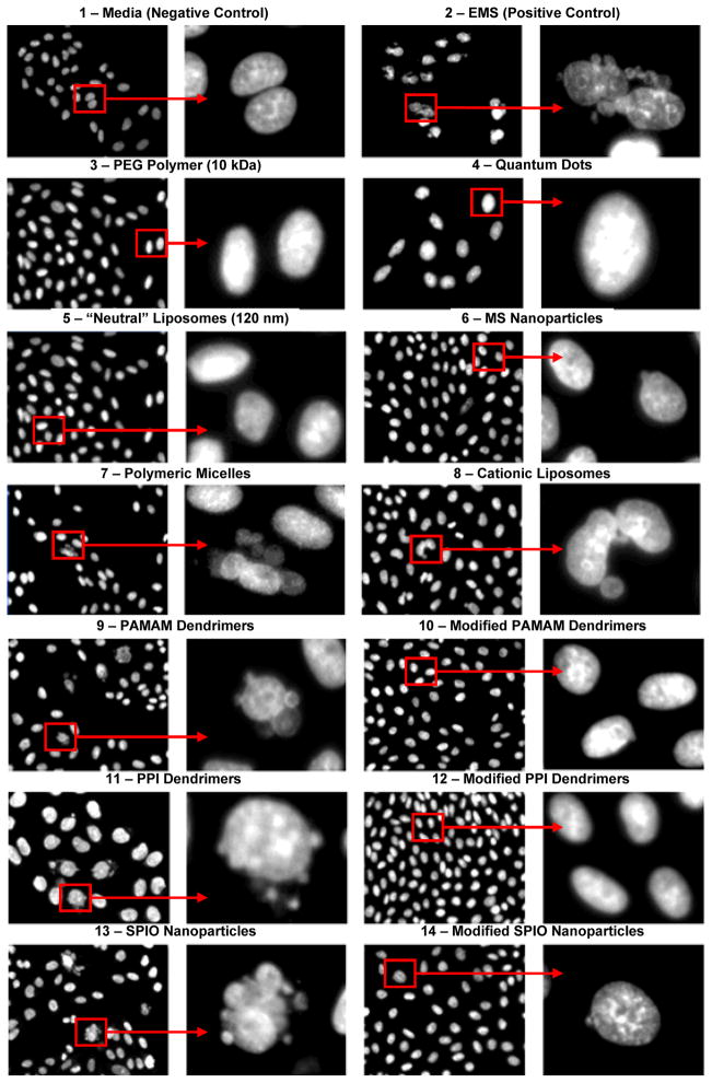
Figure 4.
Genotoxicity (formation of micronuclei) of different nanocarriers and corresponding controls. Representative fluorescence microscopy images of CHO-K1 cells incubated within 24 hours with different substances: 1 – media (negative control); 2 – ethyl methanesulfonate (EMS, positive control); 3 – 10 kDa poly(ethylene glycol) (PEG) polymer; 4 – quantum dots (QD); 5 – neutral liposomes (120 nm); 6 – mesoporous silica (MS) nanoparticles; 7 – polymeric micelles; 8 – cationic liposomes; 9 – poly(amido amine) (PAMAM) dendrimers; 10 – modified PAMAM dendrimers; 11 – poly(propyleneimine) (PPI) dendrimers; 12 – Modified PPI dendrimers; 13 – supermagnetic iron oxide (SPIO) nanoparticles; and 14 – modified SPIO nanoparticle. The cells were stained with DAPI nuclear dye. For each substance, images on the right panel show magnified cells marked by the square on the left panel.

