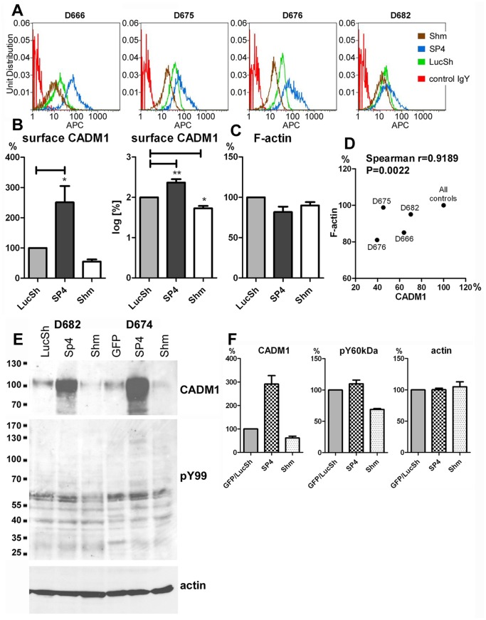
Figure 3. Downregulation of CADM1 in human lung mast cells reduced the assembly of filamentous actin and tyrosine phosphorylation.
A. HLMCs from various donors were transduced with SP4, control shRNA (LucSh) or CADM1 shRNA (Shm) viral particles, followed by fluorescent staining of surface CADM1 and F-actin. Surface CADM1 expression is shown in histograms. B. CADM1 in the SP4 and Shm groups was expressed as percentages of the control LucSh group and quantified as percentage and log-transformed percentage. (n = 5 for LucSh and SP4, n = 4 for Shm). These data were presented in Fig. 1 in [19]. C. HLMCs with modulated CADM1 were also co-stained for F-actin. The data are shown in the graph. Correlation between surface CADM1 and F-actin in LucSh- or Shm-transduced HLMCs is shown in a scatter plot with correlation parameters. E. Western blotting of protein extracts from LucSh-, SP4- and Shm-transduced HLMCs from D682 and D674, developed with Abs shown on the right. The protein bands were quantified and the data are shown in the graph.

