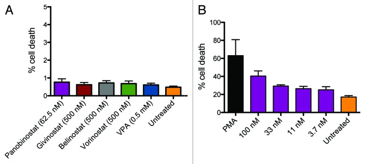
Figure 2. Toxicity in lymphocytes and U1 cells treated with HDAC inhibitors. PBMCs from healthy donors were treated with panobinostat (LBH589) (2–62.5nM), givinostat (ITF2357), belinostat (PXD101), vorinostat (SAHA) (all 62.5–500nM) and valproic acid (VPA) (0.5mM). Panel A shows cell death in lymphocytes evaluated with flow cytometry using viability stain. Only data from the highest concentration of HDAC inhibitor is shown in figure. Toxicity of panobinostat was evaluated in U1 cells using a similar experimental setup. Panel B shows cell death in U1 cells treated with panobinostat. DMSO was used as negative control and PMA as positive control.

An official website of the United States government
Here's how you know
Official websites use .gov
A
.gov website belongs to an official
government organization in the United States.
Secure .gov websites use HTTPS
A lock (
) or https:// means you've safely
connected to the .gov website. Share sensitive
information only on official, secure websites.
