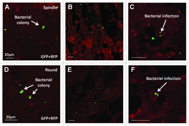
Figure 4. Confocal imaging of cancer cells infected with A1-R, expressing GFP, in vivo. Bacterial colonies were detected in both stem-like (A) and non-stem (D) tumors. Frozen section microscopy showed infiltrating A1-R in both spindle-shaped stem-like (B and C) and round non-stem cells (E and F). (C and F) are high magnification images of (B and E), respectively. Scale bars, 20 μm.

An official website of the United States government
Here's how you know
Official websites use .gov
A
.gov website belongs to an official
government organization in the United States.
Secure .gov websites use HTTPS
A lock (
) or https:// means you've safely
connected to the .gov website. Share sensitive
information only on official, secure websites.
