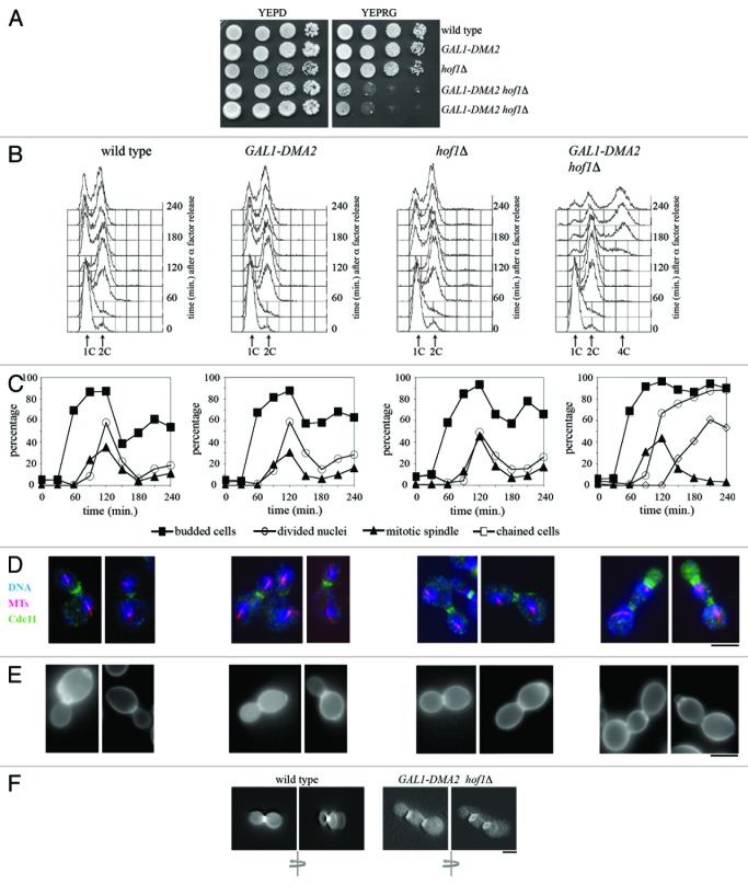
Figure 1. Dma2 excess is toxic for cells lacking Hof1.(A) Serial dilutions of stationary phase cultures of strains with the indicated genotypes were spotted on YEPD or YEPRG plates, which were then incubated for 2 d at 25 °C. (B–F) Exponentially YEPR growing cultures of wild-type, GAL1-DMA2, hof1Δ, and GAL1-DMA2 hof1Δ cells were arrested in G1 by α-factor and released from G1 arrest in YEPRG at 25 °C (time 0). At the indicated times after release, cell samples were taken for FACS analysis of DNA contents (B), and for scoring budding, chained cells, nuclear division, and mitotic spindle formation (C). (D and E) Pictures were taken 120 min after release for wild-type, GAL1-DMA2, and hof1Δ cells, or 240 min after release for GAL1-DMA2 hof1Δ cells, to show in situ immunofluorescence analysis of nuclei (DNA), mitotic spindles (MTs) and septin ring deposition (Cdc11) (D), as well as chitin deposition analysis (E) by using Calcofluor White staining. (F) Three-dimensional reconstruction of pictures taken at different Z-stacks of wild-type and GAL1-DMA2 hof1Δ cells treated as in panel (E). The arrows indicate the direction of rotation. Wild-type cells show a complete chitin disk at the bud neck (left), while GAL1-DMA2 hof1Δ cells can only assemble a chitin ring (right). Bar, 5 μm.

An official website of the United States government
Here's how you know
Official websites use .gov
A
.gov website belongs to an official
government organization in the United States.
Secure .gov websites use HTTPS
A lock (
) or https:// means you've safely
connected to the .gov website. Share sensitive
information only on official, secure websites.
