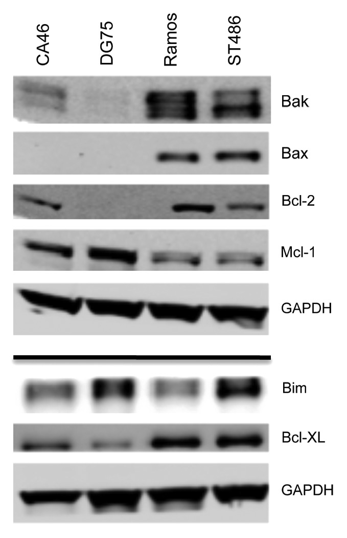
Figure 4. Expression of apoptotic proteins in Burkitt lymophoma cell lines. Whole-cell lysates were extracted from the Burkitt lymphoma lines, after which the proteins were separated by PAGE and transferred to nitrocellulose membranes. The membranes were subsequently probed with antibodies to Bak, Bax, Bcl-2, Bim, Mcl-1, and Bcl-XL. GAPDH served as a loading control. Results from one of two independent experiments are shown.
