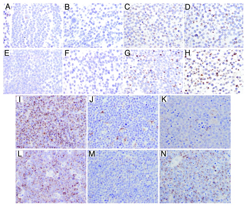
Figure 6. Immunohistochemical detection of Bak and Bax in Burkitt lymphoma cell lines and patient samples. Bak and Bax expression was examind by immunohistochemistry as outlined in the methods section. (A–D) Bak expression in Burkitt cell lines: CA46 (A), DG75 (B), Ramos (C) and ST486 (D). (E–H) Bax expression in Burkitt cell lines: CA46 (E), DG75 (F), Ramos (G) and ST486 (H). Bak (I–K) and Bax (L–N) expression in Burkitt lymphoma samples. More than 75% of tumor cells showed granular cytoplasmic expression for Bak (I) and Bax (L) in case 1 of Burkitt lymphoma; less than 25% of tumor cells were positive for Bak (J) and Bax (M) in case 11; in case 6, the tumor cells showed a discordant expression of Bak and Bax, with low expression (<25%) for Bak (K) and high expression (>75%) for Bax (N); scattered macrophages were also positive for Bak (J and K).

An official website of the United States government
Here's how you know
Official websites use .gov
A
.gov website belongs to an official
government organization in the United States.
Secure .gov websites use HTTPS
A lock (
) or https:// means you've safely
connected to the .gov website. Share sensitive
information only on official, secure websites.
