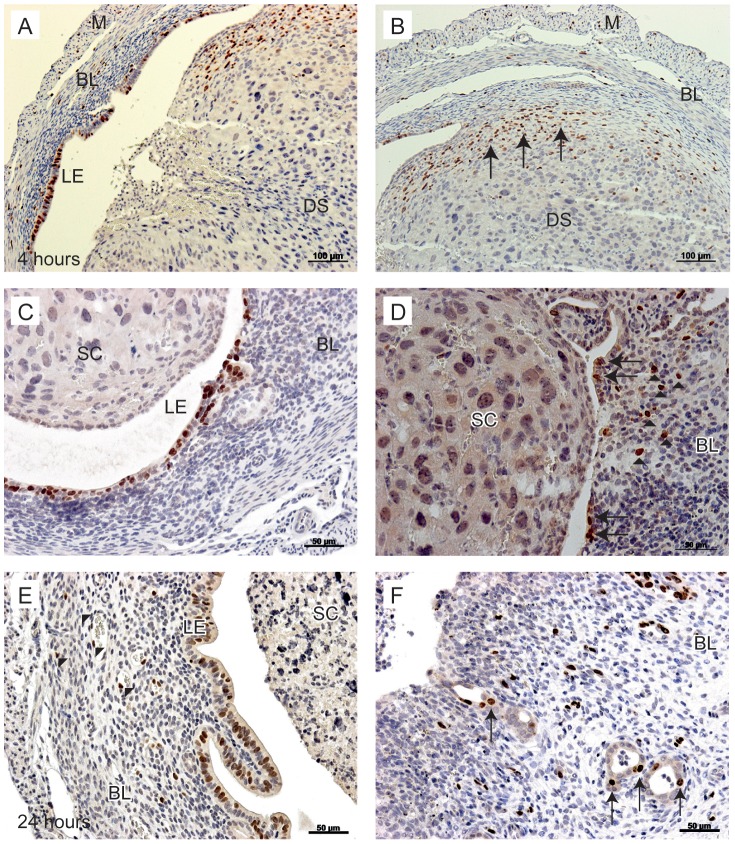
Figure 3. Proliferation of uterine cells between 4 and 24 hours after P4 withdrawal.
To identify proliferating cells, animals were injected with BrdU 90 minutes prior to tissue recovery. A; Proliferating luminal epithelial cells detected in tissues 4 hours after progesterone withdrawal. B; In the same tissue, stromal cells in the basal layer are positive for BrdU (arrows). C; At 12 hours, luminal epithelial cells were positive for BrdU, no BrdU positive cells were identified in the shed cell mass. D; In the same tissue, stromal cells close to the luminal edge were positive for BrdU (arrowheads), new epithelial cells were identified lining the lumen in an area of tissue where the decidualised tissue had shed (arrows). E; At 24 hours after withdrawal, endothelial cells were positive for BrdU (arrowheads), the intact luminal epithelium was also positive for BrdU. F; In another sample at 24 hours, the stromal compartment was exposed to the lumen (arrowheads); note stromal cells positive for BrdU are interspersed throughout the basal layer and evidence of glandular epithelial cell proliferation was also detected (arrows). BL; Basal layer, LE; luminal epithelium, DS; decidualised stromal cells, M; myometrium, SC; shed cells. Scale bars are equal to 100 µm or 50 µm where indicated.

