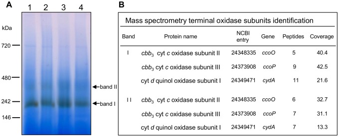
Figure 3. Identification of cytochrome c oxidase(s) in S. oneidensis MR-1 membranes under aerobic and microaerobic conditions.
A. In-gel detection of cytochrome c oxidase activity in solubilized membranes on BN-gel. Membrane proteins were prepared from S. oneidensis MR-1 cells grown aerobically (lanes 1 and 2) or microaerobically (lanes 3 and 4) and harvested during the exponential (lanes 1 and 3) or the stationary (lanes 2 and 4) phase of growth. 130 µg of total proteins were loaded on a 5–15% polyacrylamide gel. The major brown band (band I) and the faint brown band (band II) of activity are indicated by arrows. B. Terminal oxidases subunits identified by ESI-Q-ToF mass spectrometry. Data correspond to identified proteins in bands I and II from exponentially grown cells under aerobic conditions (panel A, lane 1). Table heading: Band, roman figures refer to the protein bands from the BN gel shown in panel A; Protein name, name in NCBI database; NCBI entry, accession number; Peptides, number of unique peptides detected; Coverage, protein sequence coverage by the matching peptides (in %). Cyt: cytochrome.

