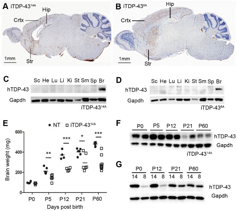
Figure 1. Expression of human TDP-43 in iTDP-4314A and iTDP-438A mice in the postnatal period.
Immunohistochemical detection of hTDP-43 expression in cortex (CTX), hippocampus (HIP) and striatum (STR) in iTDP-4314A (A) and iTDP-438A (B). Western analysis of organs demonstrated specificity of hTDP-43 expression to the brain in both iTDP-4314A (C) and iTDP-438A (D) (SC = spinal cord, He = heart, Lu = lung, Li = liver, Ki = kidney, St = stomach, SM = skeletal muscle, Sp = spleen, Br = brain). (E) Brain weight measurement of non-transgenic (NT) and iTDP-4314A mice at postnatal stages until 2 months of age (P60) (*p<0.05, **p<0.01, *** p<0.001, unpaired two tailed T-test). (F) Expression of hTDP-43 at indicated postnatal time points for iTDP-4314A. (G) Expression of hTDP-43 at indicated postnatal time points for iTDP-4314A (14) compared to iTDP-438A (8).

