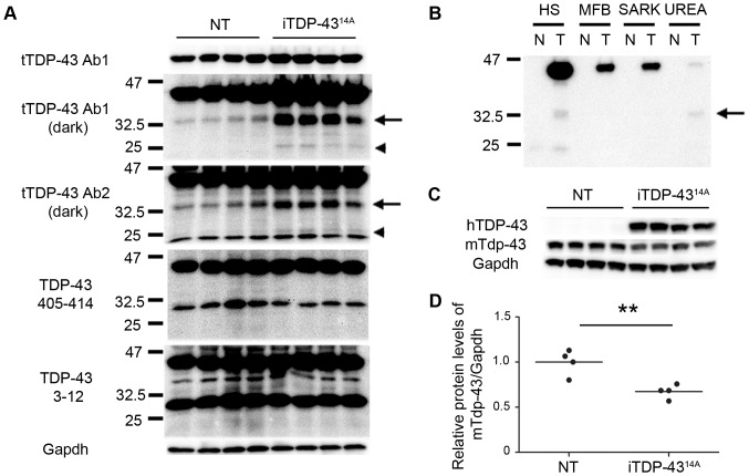
Figure 3. Biochemistry of iTDP-4314A brain lysates at P5.
(A) Western blotting using two antibodies to total TDP-43 (tTDP-43 Ab1 and tTDP-43 Ab2) demonstrated increased levels of low molecular weight species at 35 kDa (arrow) and 25 kDa (arrowhead) in iTDP-4314A mice relative to NT mice. These species were not observed using antibodies to the C-terminus (405–414) or N-terminus (3–12) of TDP-43. (B) Western blot analysis of high salt (HS), myelin floatation buffer (MFB), sarkosyl (SARK) and urea fractions using antibody to human TDP-43. Note that human TDP-35 (arrow) is present in the urea fraction but is absent from MFB and SARK fractions, N = non-transgenic, T = iTDP-4314A. (C) Antibody to murine Tdp-43 demonstrated reduction of mTdp-43 in brain compared to NT mice. (D) Quantification of blot in (C), **p<0.01, unpaired two tailed t-test.

