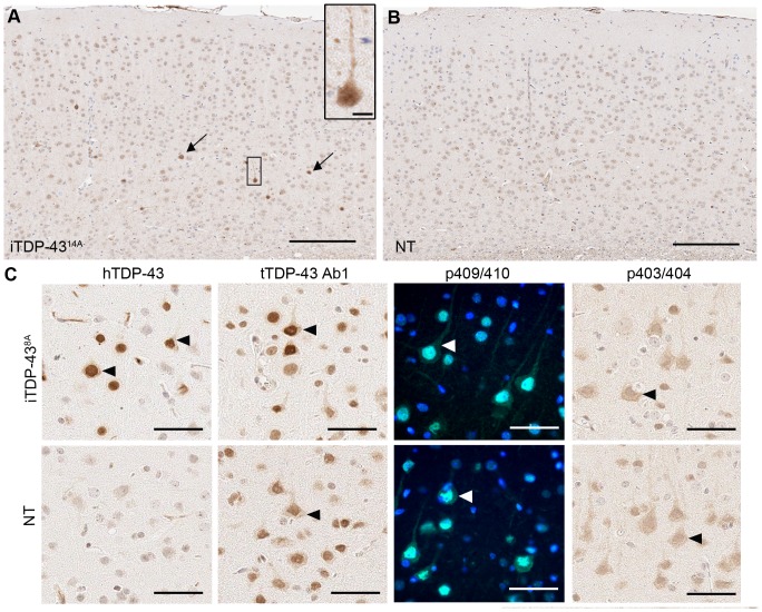
Figure 5. Neuropathology of 25 month old iTDP-438A mice.
Immunohistochemical detection of ubiquitin revealed rare cells bearing increased ubiquitin staining in the cortex of iTDP-438A mice (arrows, A) that was absent in NT animals (B, scale bar = 200 µm). Staining was detected in both nucleus and cytoplasm of affected cells (inset in A, scale bar = 10 µm). (C) In iTDP-438A animals hTDP-43 was predominantly nuclear, some cells displaying cytoplasmic localization without aggregation. Cytoplasmic localization was observed in NT and iTDP-438A mice using antibodies to total TDP-43 (tTDP-43 Ab1) and phosphorylated forms of TDP-43 (p403/404, p409/410).

