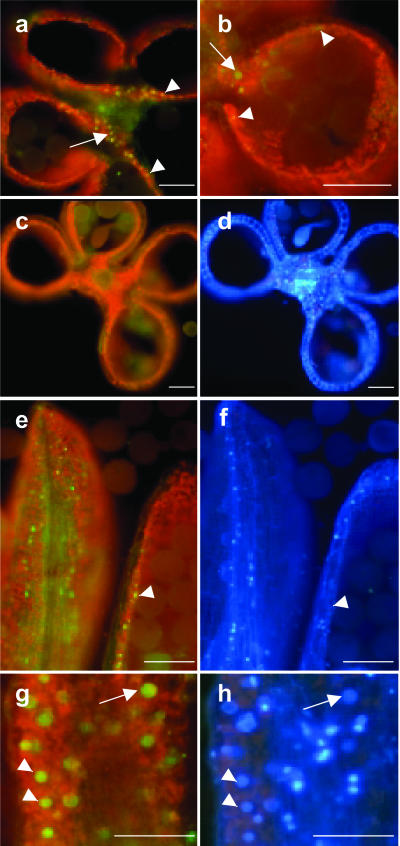
Figure 6.
ZmMADS2-GFP expression in cross and longitudinal sections of anthers from transgenic maize plants at 2 d before anthesis. a, Cross section of a late stage VIII anther of a transgenic maize line expressing the ZmMADS2-GFP-fusion protein under control of the ZmMADS2 promoter (Fig. 3) in connective tissue (arrow) and endothecial cells (arrowheads). b, Close-up of an anther locule; arrow marks nucleus in connective tissue and arrowheads in endothecial cells. c, WT anther of the same stage lacking green fluorescence. d, DAPI staining of c. e and g, Longitudinal section to show expression of the ZmMADS2-GFP-fusion protein in nuclei of endothecial cells. e, Expression of the fusion protein is not restricted to the region of unopened anterior pore. Note that nuclei showing bright GFP-fluorescence display only faint DAPI signals. g, Close-up of a longitudinal section showing expression of the ZmMADS2-GFP fusion protein in connective tissue. f and h, DAPI staining of images shown in e and g. Note that the stronger the GFP fluorescence, the weaker the DAPI signal. Arrowheads point to strong GFP fluorescence and weak DAPI staining; arrow points toward stronger GFP fluorescence and weaker GFP signals. Bars = 100 μm.

