Figure 7.
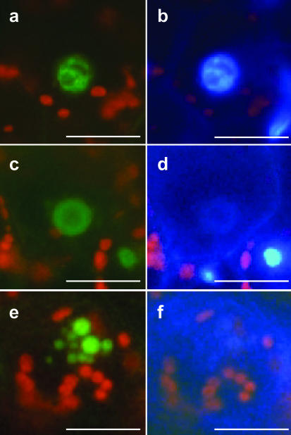
ZmMADS2 marks degrading nuclei of endothecial cells during anther maturation. a, ZmMADS2-GFP fluorescence of an intact nucleus within an endothecial cell. b, DAPI staining of the image shown in a. Note that the nucleus shows compartmentation. c, The ZmMADS2-GFP-fusion protein is visible, whereas most DNA is degraded. The structure of the nucleus is lost. d, DAPI staining of c. Few intact DNA are left. e, ZmMADS2-GFP is detectable in apoptotic bodies of an endothecial nucleus. f, DAPI staining of e. DNA is almost completely degraded. Bars = 10 μm.

