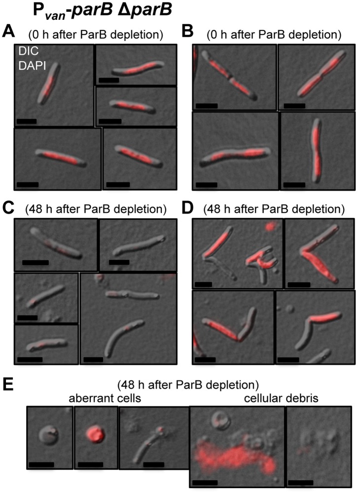
Figure 4. ParB is involved in chromosome partitioning in M. xanthus.
Merged DIC and fluorescence images of cells from a Pvan-parB ΔparB (MR2472) culture in the presence of vanillate (permissive conditions) (A) and (B), and after 48 hours in the absence of vanillate (restrictive conditions) (C), (D) and (E). The cultures were stained with DAPI for viewing chromosomal DNA by fluorescence, shown in red. Black scale bars represents 5 µm. (A) Non-dividing cells with DNA. (B) Dividing cells with DNA. (C) Non-dividing cells without DNA. (D) Dividing cells with DNA only in one compartment. (E) Rounded cells without DNA (first from left) and with DNA (second from left), broken cell without DNA (middle), extracytoplasmic DNA (second right) and cellular debris (first right). The mean of the results from three independent experiments and the standard deviations are shown in Table 1.

