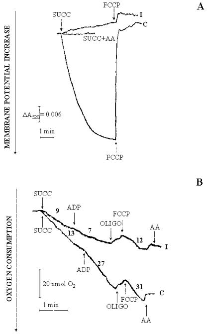
Figure 12.
Generation of ΔΨ and oxygen uptake by mitochondria isolated from control and PCD cells arising from externally added succinate. A, Mitochondria isolated from control and 4-h PCD cells (0.5 mg of protein) were incubated at 25°C in 2 mL of the medium reported in “Materials and Methods” with continuous measurement of the safranin O A520. At the arrows, succinate (5 mm) and FCCP (1.25 μm) were added. When present, antimycin (AA; 2 μg) was added 1 min before succinate addition. B, Mitochondria from control and 4-h PCD cells (0.5 mg protein) were incubated at 25°C in 1.5 mL of the respiratory medium reported in “Materials and Methods,” and oxygen uptake was measured polarographically as a function of time. At the arrows, 5 mm succinate, 0.5 mm ADP, 2.5 μm oligomycin (OLIGO), 1.25 μm FCCP, and 2 μg AA were added. The rate of oxygen consumption is expressed as nanomoles of O2 per minute per milligram of mitochondrial protein.

