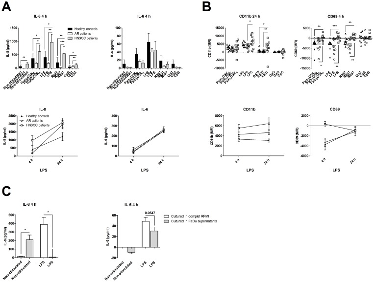
Figure 2. Increased activation of polymorphonuclear leukocytes (PMN) in patients with head and neck squamous cell carcinoma (HNSCC).
Blood was obtained from healthy controls (n = 10), patients with an ongoing seasonal allergic rhinitis (AR; n = 11) and patients with HNSCC (ELISA n = 9; FACS n = 10). PMN were isolated and cultured in the presence or absence of Pam3CSK4 (1 µg/ml), LPS (1 µg/ml), R837 (5 µg/ml) or CpG (0.3 µM) for 4 and 24 h. (A) The cell free supernatants were then analyzed for IL-6 and IL-8 with ELISA, (B) and the CD16 positive cells were investigated for the expression of CD11b and CD69 using flow cytometry. The results are presented as the non-stimulated value minus the TLR stimulated values. Grey colored samples were analyzed on a BD LSRFortessa, whereas the rest of the samples were investigated on a Beckman Coulter Navios flow cytometer. (C) PMN from healthy individuals (n = 9) were incubated with or without culture medium supernatant from the HNSCC cell line FaDu, and stimulated with or without 1 µg/ml LPS for 4 h, and analyzed for IL-6 and IL-8 secretion with ELISA. The basal cytokine levels in the media were subtracted from the concentrations present in the PMN cultures. MFI = mean fluorescence intensity; *p≤0.05; **p≤0.01; ***p≤0.001.

