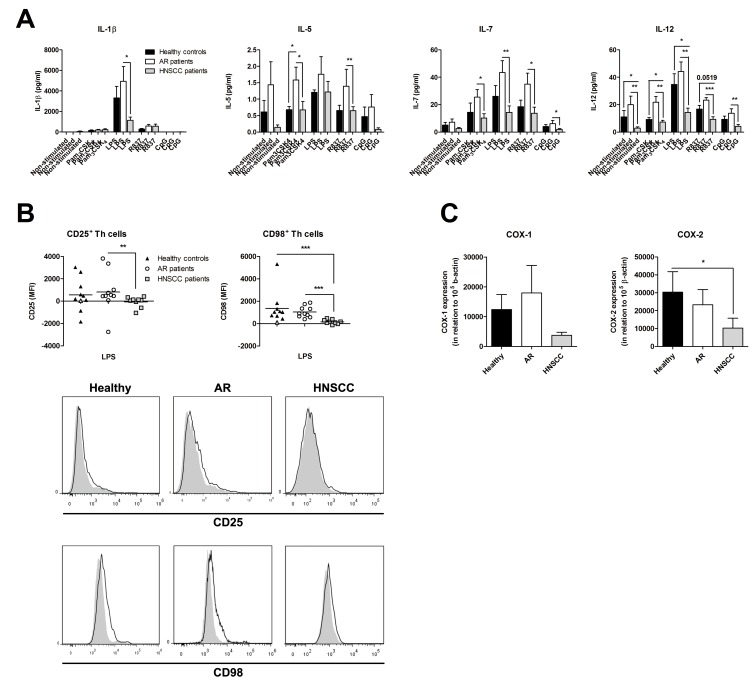
Figure 3. Decreased peripheral blood mononuclear cells (PBMC) activation in patients with head and neck squamous cell carcinoma (HNSCC).
PBMC were isolated from blood collected from healthy controls (n = 10), patients with an ongoing seasonal allergic rhinitis (AR; n = 11) and patients with HNSCC (n = 9), and cultured with or without Pam3CSK4 (1 µg/ml), LPS (1 µg/ml), R837 (5 µg/ml) or CpG (0.3 µM) for 24 h. (A) Thereafter, the supernatants were analyzed for the secreted cytokine profile with Luminex Multiplex Immunoassay, 4 out of 17 investigated cytokines are shown, (B) and the cells were examined for the expression of CD25 and CD98 on CD4 positive T helper (Th) cells with flow cytometry. The results are presented as the non-stimulated value minus the TLR stimulated values. Grey colored samples were analyzed on a BD LSRFortessa, whereas the rest of the samples were investigated on a Beckman Coulter Navios flow cytometer. One representative histogram from the healthy controls, AR patients and HNSCC patients is displayed. The open histograms represent LPS stimulated cells, and the light grey histograms are denoting the non-stimulated samples. (C) RNA was extracted from peripheral PBMC, isolated from healthy individuals (n = 8), AR patients (n = 10), and HNSCC patients (n = 7), made into cDNA, and thereafter analyzed for COX-1 and COX-2 mRNA expression with real-time PCR. Data are given in relation to the housekeeping gene β-actin, 100 000×2−ΔCt, and presented as mean ± SEM. MFI = mean fluorescence intensity; *p≤0.05; **p≤0.01; ***p≤0.001.

