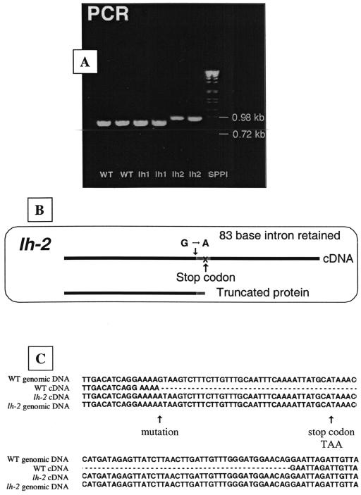
Figure 6.
The nature of the lh-2 mutation. A, Agarose (2% [w/v])/Tris-acetate EDTA (TAE) electophoresis gel of PCR products with the same primers from wild-type, lh-1, and lh-2 cDNA. B, Schematic diagram of lh-2 mutation. C, Comparison of genomic and cDNA sequence from wild type and lh-2 to illustrate the lh-2 genetic lesion.

