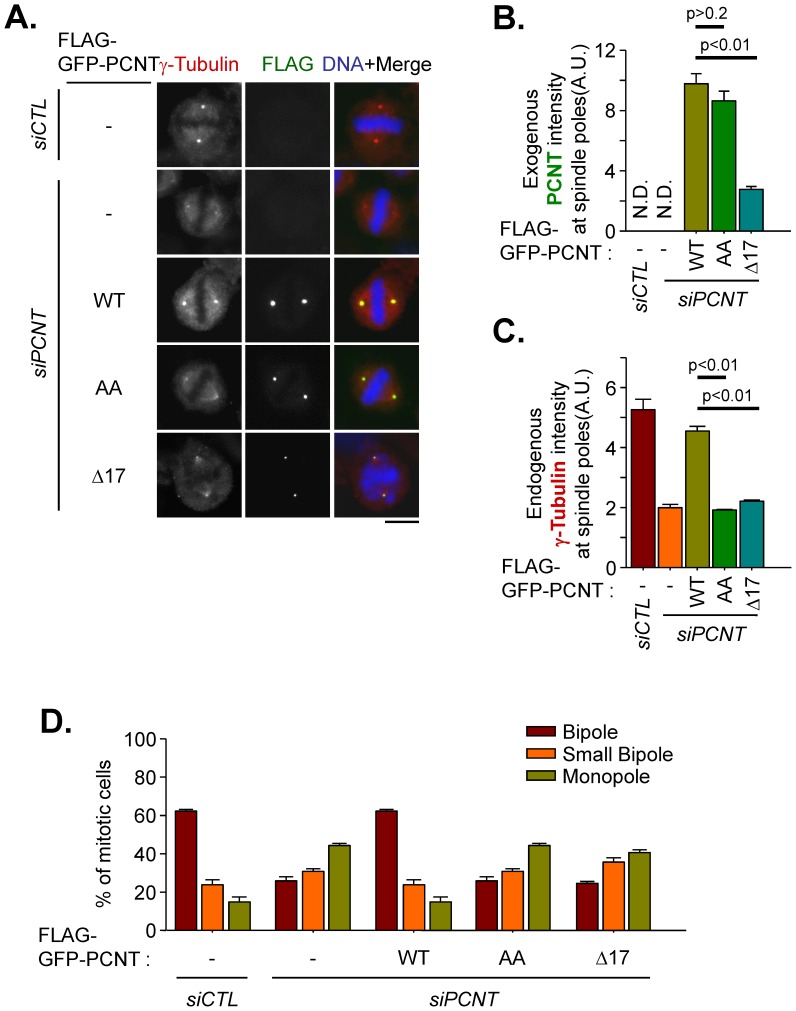
Figure 4. Spindle defects in cells rescued with the pericentrin mutant.
(A–C) Pericentrin-depleted HeLa cells were rescued with FLAG-GFP-tagged PCNT (WT), PCNT1235,1241AA (AA) and PCNTΔ2390–2406 (Δ17). The cells were treated with RO3306 for 16 h and subsequently removed for 40 min to allow accumulation of mitotic cells. The cells were coimmunostained with γ-tubulin (red) and FLAG (green) antibodies. Scale bar, 10 µm. The intensities of ectopic pericentrin (B) and endogenous γ-tubulin (C) at the spindle poles were quantified in more than 40 cells per group in three independent experiments. Error bars, SEM. The paired t-test was performed with p value indicated. (D) PCNT-depleted HeLa cells were rescued with FLAG-GFP-tagged PCNT (WT), PCNT1235,1241AA (AA) and PCNTΔ2390–2406 (Δ17). The cells were treated with RO3306 for 16 h and released with STLC for additional 1 h to arrest the cells at prometaphase. Then, the cells were washed and re-incubated with MG132 for 1.5 h to avoid the cells progress to anaphase. The cells were coimmunostained with FLAG and NuMA antibodies. The phenotype of the bipolar spindle was categorized as bipole, small bipole or monopole based on the NuMA staining patterns.

