
Figure 3.
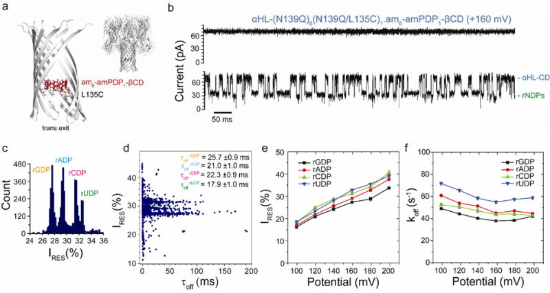
rNDP interaction with the αHL-(N139Q)6(N139Q/L135C)1.am6-amPDP1-βCD pore. (a) Structure of the αHL-(N139Q);6(N139Q/L135C)1.am6-amPDP1- βCD pore (cartoon view). The enlarged view shows a close-up of the β barrel with two subunits omitted. The cyclodextrin (am6-amPDP1-βCD) was covalently attached through a disulfide bond to position 135 in one of the seven subunits, which had been mutated to Cys (red, sticks model)21(b) Single-channel recording from the αHL-(N139Q)6(N139Q/L135C)1.am6-amPDP1-βCD pore showing continuous rNDP detection. (c) Corresponding residual current (IRES%) histogram. Data were acquired in 1 M KCl, 25 mM Tris-HCl, pH 7.5, at +160 mV in the presence of 10 μM rGDP, 10 μM rADP, 10 μM rCDP and 10 μM rUDP (all cis). The results displayed are from a typical experiment. (d) Scatter plot showing IRES% and dwell times (τoff) for rNDP binding events to the αHL-(N139Q)6(N139Q/L135C)1.am6-amPDP1-βCD pore as seen in the current trace in ‘b’. (e) Variation of residual currents (IRES%) with applied potential for each rNDP detected with the αHL-(N139Q)6(N139Q/L135C)1.am6-amPDP1βCD pore. (f) Variation of k Values of koff with applied potential for each rNDP detected with the αHL-(N139Q)6(N139Q/L135C)1.am6-amPDP1-βCD pore. Values of koff were determined by using koff = 1/τoff where τoff is the mean dwell time of the rNDP in the pore.