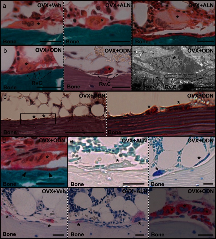
Fig. 2.
Histological appearance of osteoclasts in odanacatib (ODN)– and alendronate (ALN)–treated ovariectomized (OVX) rabbits. a Masson-Goldner trichrome staining shows the appearance of osteoclast profiles (asterisks) attached to the vertebral trabecular bone surface from a vehicle-treated (left panel) and an ALN-treated (middle panel) OVX rabbit. Osteoclast profiles frequently appeared away from the bone surface in ALN-treated OVX rabbits as shown by the presence of a giant osteoclast (asterisk) above bone forming osteoblasts (right panel). b Examples of osteoclast profiles (asterisks) detached from the bone surface by mononucleated reversal cells (Rv.C, arrows) in an ODN-treated OVX rabbit as they appear using Masson-Goldner trichrome staining (left panel), TRAP activity staining (middle panel), and electron microscopy (right panel). c TRAP-positive osteoclast profiles (asterisks) attached to a bone surface without visible broken lamellae. The framed area in the left panel is enlarged in the right panel. d Example of an osteoclast profile (asterisk) with demineralized collagen below (arrowheads), visualized by Masson-Goldner trichrome staining. e Toluidine blue staining showing the absence and presence of intracellular vesicles in osteoclast profiles (asterisks) in the vertebral trabecular bone from an ALN-treated (left panel) and an ODN-treated (right panel) OVX rabbit. f Immunohistochemical staining showing elevated intracellular levels of cathepsin K in trabecular osteoclasts (asterisks) of ODN-treated OVX rabbits (right panel) compared to ALN-treated (middle panel) and vehicle-treated (left panel) OVX rabbits. Scale bar = 50 μm, except for b, right panel = 10 μm and c, left panel = 200 μm

