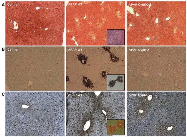
Figure 2.
(A) Representative liver sections of untreated control mice or animals treated for 6 h with 200 mg/kg acetaminophen (APAP) and stained with H&E. (B) DNA fragmentation as evaluated by the TUNEL assay in mice treated with APAP for 6 h. (C) Nitrotyrosine staining as an indicator of peroxynitrite formation in mice treated with APAP for 6 h. Inserted images of serial liver sections from wild type animals treated with APAP indicate that the centrilubular location of cell necrosis correlates with the area of TUNEL and nitrotyrosine staining.

