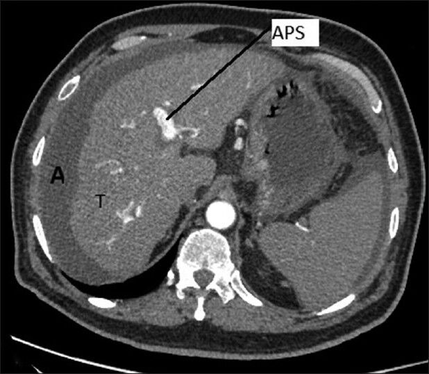
Figure 1.

Arterial phase of triple phase contrast enhanced computer tomography revealed mass lesion of 12 cm × 15 cm size in segments 6 and 7 of liver. tumor enhancement (T) in arterial phase and portal vein is visible on arterial phase (arterioportal shunt) and ascites (A)
