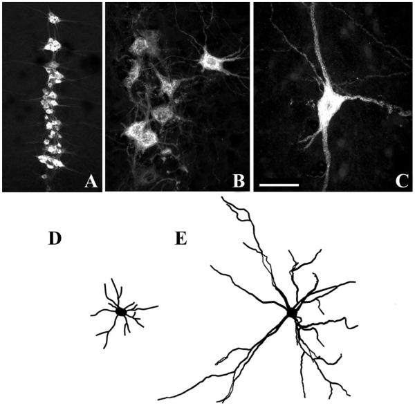
Fig. 3.
Confocal photomicrographs displaying phrenic motoneurons that were retrogradely labeled by DIAm injection of cholera toxin B-fragment. Motoneurons are shown at postnatal day 10 (P10, A and B) and in the adult rat (C). Neurolucida camera tracings are shown in D and E for P10 and adult motoneurons, respectively. Note differences in cell size (B and C) and dendritic arborization (D and E). Bar indicates 200 μm (A) and 50 μm (B and C).

