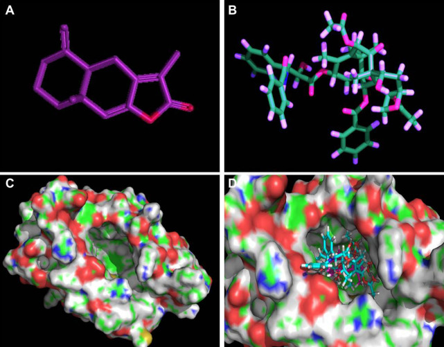
Figure 4. Molecular model of docking of paclitaxel and AO-I to the molecular model of MD-2.
(A and B). 3D structures of AO-I and paclitaxel were drawn as a ball-and-stick representation: AO-I in purple, paclitaxel in blue-green. (C). 3D structure of human MD-2. Protein surface showing hydrophobic and hydrophilic properties. Green and red represent hydrophobicity and hydrophilicity, respectively, and the surface center of MD-2 with a potential binding-pocket (iron-gray); (D). AO-I binding to the hydrophobic pocket of MD-2, which partially overlaps with the binding site of paclitaxel.

