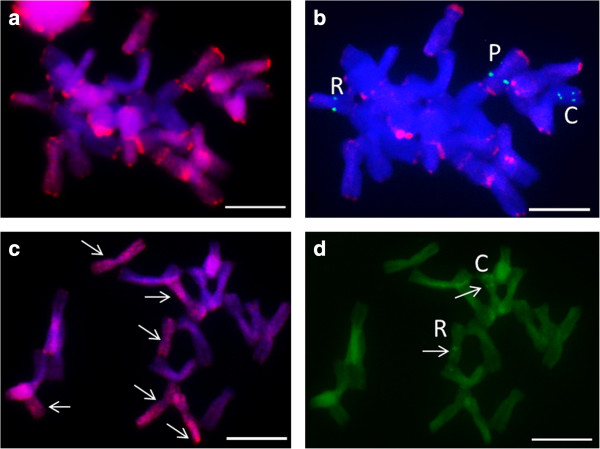
Figure 4.
Genomic in situ hybridisation (GISH); a, c) and subsequent 5S rDNA mapping (b, d) to mitotic metaphase chromosomes of Allium × cornutum. (a) GISH with genomic DNA of A. pskemense (red) and A. cepa as blocking DNA, incomplete metaphase plate; (b) 5S rDNA (green) localisation in the same chromosomal spread (c) GISH with genomic DNA of A. roylei (red) and A. cepa as blocking DNA, incomplete metaphase plate; (d) 5S rDNA (green) mapping in the same chromosomal spread. The letters C, R, and P indicate chromosomes carrying the 5S signal and belonging to the three different genomes (due to insufficient washing of the genomic probe, red subtelomeric signals (b) remained visible in majority of the chromosomes). Scale bar = 10 μm. A few of the nuclei (top left and right corners) visible in (a) were lost during reprobing and are not visible in (b).

