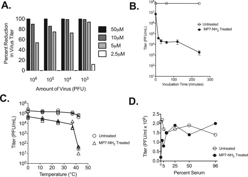
Fig. 1.
MP7-NH2 inactivates VSV. (Panel A) Increasing amounts of purified VSV were incubated with increasing concentrations of MP7-NH2 as indicated. Reactions were performed at room temperature in a total volume of 500 μl with serum free DMEM as the diluent. After a 30 min incubation cultures were serially diluted, and applied to monolayers of BHK cells to detect residual infectious virus. Plaques were counted 48 h later. Bars on graph represent the percent reduction in virus titer when the untreated and treated samples were compared. (Panel B) 107 PFU of VSV was incubated with 50 μM MP7-NH2 at room temperature for the indicated amounts of time. Graph represents the average ±SEM of three experiments performed independently. (Panel C) 105 PFU of VSV was incubated with 10 μM MP7-NH2 at the indicated temperatures for 30 min. Graph shows results of three experiments performed independently. (Panel D) 106 PFU of VSV was incubated with 10 μM MP7-NH2 at room temperature for 30 min in the presence of the indicated concentration of non-heat inactivated fetal calf serum. Panel D shows one representative graph from four studies performed independently.

