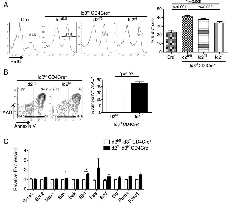
Figure 3.
Conditional knockout of both Id2 and Id3 impairs the proliferation and survival of Vδ6.3+ γδ T cells. (A) In the neonatal thymus, Vδ6.3+ γδ T cells from Id3f/f CD4Cre+ mice are more highly proliferative than the Cre− controls as shown by BrdU incorporation assay, regardless of their Id2 genotype. However, cells from the Id2f/f Id3f/f CD4Cre+ mice show a small but significant decrease in BrdU+ cell percentage compared to those from Id2B/BId3f/fCD4Cre+ mice. n=3 for each group. (B) Vδ6.3+ γδ T cells were sorted from the thymus of neonatal mice and cultured for 24 hours. Id2f/f Id3f/f CD4Cre+ cells showed increased cell death by 7AAD and Annexin V staining compared to Id2f/B Id3f/f CD4Cre+ cells. n=3 in each group. (C) QPCR analysis of a panel of cell death-related genes showed that Id2f/f Id3f/f CD4Cre+ Vδ6.3+ γδ T cells express more mRNA of pro-apoptotic genes Bim and Bax. n=3 in each group. *p<0.05. All error bars indicate SD.

