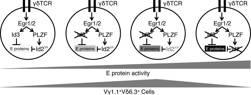
Figure 6.
A schematic diagram of Vγ1.1+Vδ6.3+ γδ T cell developmental control by Id2 and Id3. In the developing thymus, γδ T cells that express the Vγ1.1 and Vδ6.3 TCR segments receive strong TCR signaling, up-regulating Id2 and Id3 through Egr1/2 and PLZF. The Id proteins inhibit activity of E proteins, affecting the survival and proliferation of Vγ1.1+Vδ6.3+ γδ T cells. When Id3 is present, and Id2 is expressed from a more active allele, such as the one from the 129 genetic background (Id2s, “strong”), E protein activity is very low and Vγ1.1+Vδ6.3+ γδ T cell population size is small. If Id3 is absent, and Id2 is expressed from a less active allele, such as the one from the B6 background (Id2B, “B6”), E protein activity becomes higher and the Vγ1.1+Vδ6.3+ γδ T cells expand dramatically. However, if both Id2 and Id3 are completely absent, E protein activity becomes too high and again impairs the survival and proliferation of Vγ1.1+Vδ6.3+ γδ T cells, limiting its population size.

