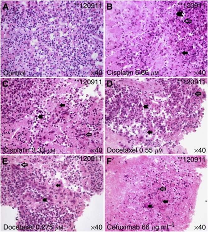
Figure 3.
Nuclear fragmentation after treatment. (A) HE-stained untreated control kept in vitro for 6 days. Note the euchromatic nuclei and an absence of nuclear fragments. (B–F) Slices treated with cisplatin 6.66 μM (B), cisplatin 3.33 μM (C), docetaxel 0.55 μM (D), docetaxel 0.275 μM (E) or cetuximab 66 μg ml−1 (F) exhibit fragmented nuclei (arrow, filled), pycnotic alterations (arrow, not filled) and cellular polymorphisms (arrow head) as hallmarks of apoptosis, which are most prominent after exposure to high dose of cisplatin (B) and cetuximab (F). Note that some regions seem to be depleted from cells.

