
Figure 1.
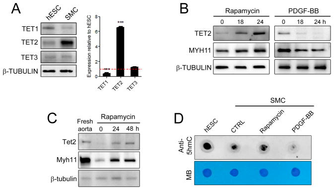
TET2 is associated with the differentiated SMC state. (A) Western blot showing protein levels of TET1, TET2 and TET3 in human coronary artery smooth muscle cells (SMC) compared to human embryonic stem cells (hESC). Corresponding relative mRNA levels are shown on the right (qPCR). Relative expression of hESC is set to 1.0 and denoted by the red line. Data are presented as mean ± SD for three independent experiments. ***P<0.001. (B) Western blot showing TET2 and MYH11 levels in hCASMC following 50 nM rapamycin or 5 ng/ml PDGF-BB treatment. (C) Western blot comparing Tet2 and Mhy11 levels from freshly isolated mouse aorta tissue and cultured cells. (D) Dot blots for global 5-hmC expression using genomic DNA isolated from hESC and hCASMC treated as in (B) for 48 hours. Methylene blue (MB) staining demonstrates equal loading.