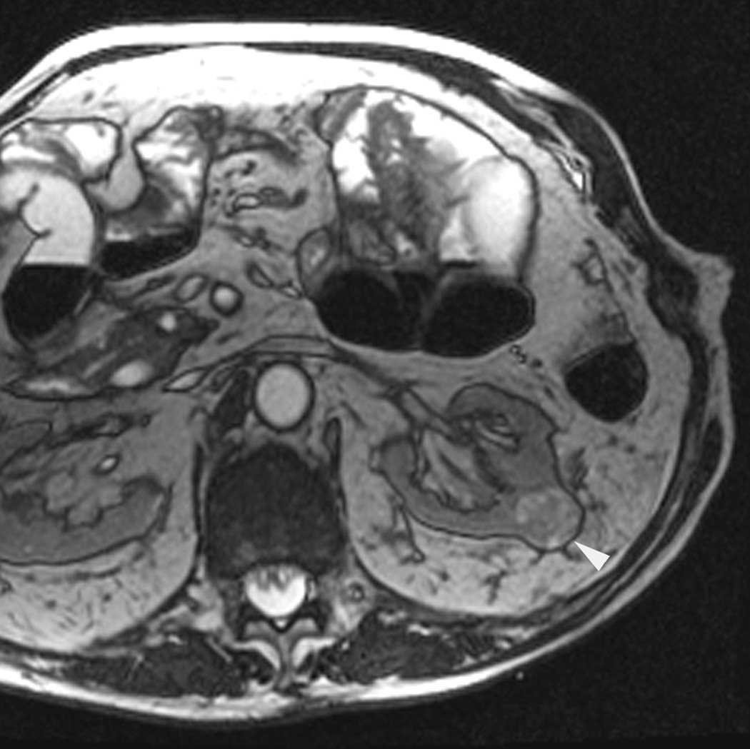
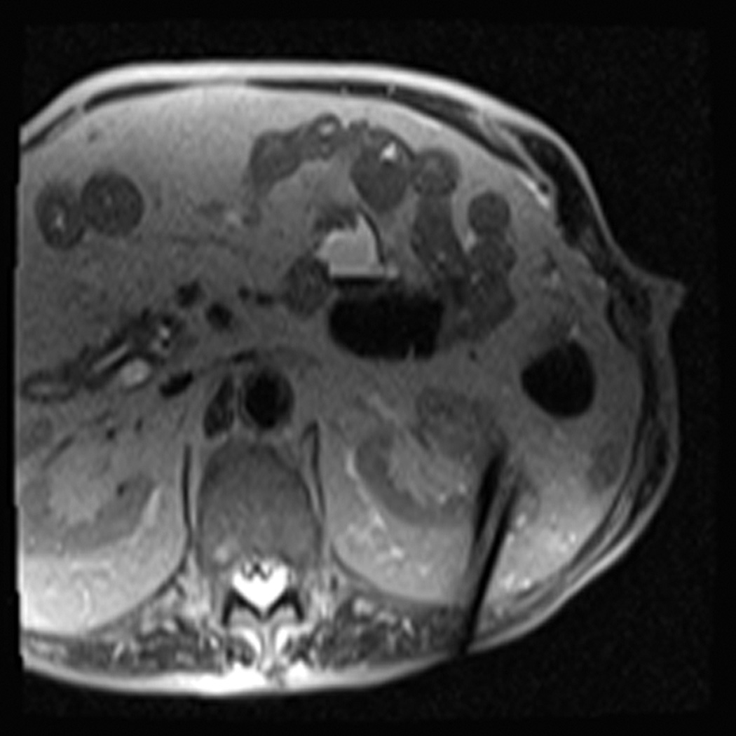
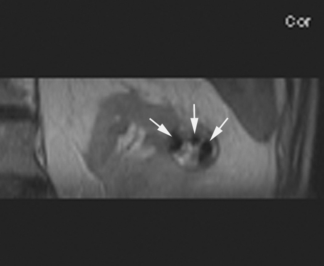
Figure 2.
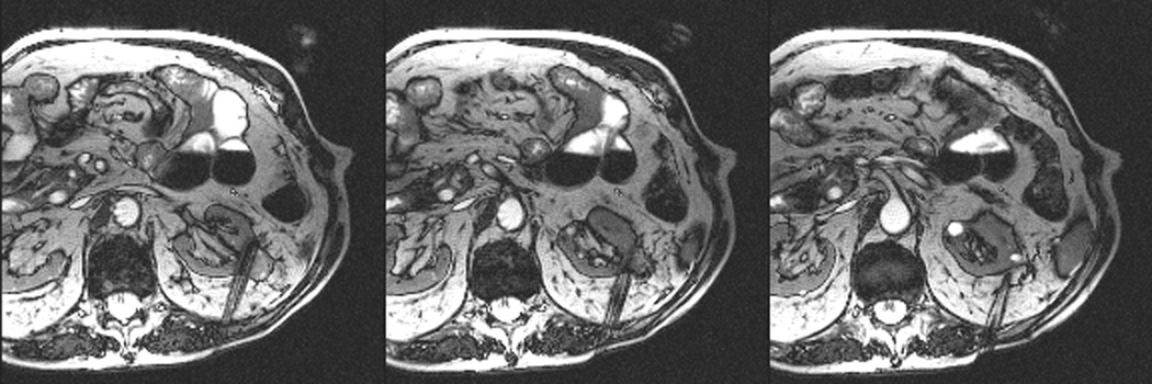
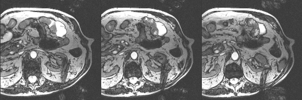
An 80 year old man was found to have a 0.8 cm mass in the left kidney. He was initially managed by active surveillance. Repeat imaging showed enlargement of the tumor to 2.8 cm. Biopsy showed renal cell carcinoma, clear cell type, Fuhrman nuclear grade 2. He was referred for image-guided ablation. Axial T2-weighted (bSSFP) MRI of the abdomen in prone position (a) shows a hyperintense mass in the lateral aspect of the left kidney. The first cryoprobe was inserted into the medial border of the mass (b) under real-time MRI guidance (prtototype bSSFP sequence). Three parallel sequentially acquired images (4 mm thick with 1 mm gap) are displayed and updated at a rate of < 1 second per slice (b). The center image is the center slice, the image to the left is the inferior plane, and the image on the right is the superior plane. These three parallel images help monitor any deviation of the probe from its intended course. The operator can adjust the angle as needed and continue to insert the probe under real time MRI guidance. Another set of images (c) shows placement of the second probe in the lateral aspect of the tumor. Susceptibility induced signal losses degraded the quality of the bSSFP acquisition (c). Axial images were obtained using a HASTE sequence to better delineate the position of the probes (d). After insertion of the third probe under real-time guidance, a 3-D HASTE sequence was performed. Reconstructed coronal image (e) shows the distribution of the probes within the tumor (arrows). Note that the scanner displays the image in the default radiological coordinates (ventral surface at top of image) despite the fact that the patient is placed in prone position.

