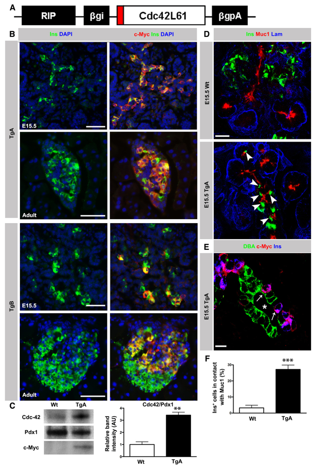
Fig. 2.
Expression of constitutively active Cdc42 in pancreatic β cells impairs delamination. (A) Schematic of the transgenic construct. A c-Myc tagged (red) constitutively active form of Cdc42 cDNA was cloned under the rat insulin promoter (RIP) along with rabbit β-globin intron (βgi) and polyA (βgpA). (B) Double immunostaining of E15.5 and adult (8-week-old) Tg pancreas sections with antibodies against c-Myc (red) and Ins (green) together with DAPI (blue). In TgA, >90% of Ins+ cells co-expressed Ins and c-Myc. In the TgB line, co-expression of c-Myc in Ins+ cells varied between 50 and 75%. In both Tg lines, expression of the transgene was restricted to Ins+ cells. (C) Immunoblot analysis of Cdc42 protein expression in 8-week-old Wt and TgA islets with Cdc42, c-Myc and Pdx1 antibodies. Quantification of the Cdc42 band intensity (normalized to Pdx1) showed a threefold overexpression in TgA islets compared with Wt (n=3, **P=0.0018). (D) Triple immunostaining of sections from E15.5 Wt and TgA pancreas with antibodies against Ins (green), Muc1 (red) and laminin (Lam; blue). In the Wt, the majority of Ins+ cells had delaminated and cells were distributed outside the duct epithelium. A significant number of TgA Ins+ cells remained in contact with Muc1 (arrowheads). (E) Triple immunostaining of sections from E15.5 TgA pancreas with antibodies against the duct cell marker DBA (green), c-Myc (red) and Ins (blue). TgA Ins+ cells (arrows) were distributed between DBA+ cells. Asterisk indicates lumen. (F) Quantification of the Ins+ cell number facing the duct lumen (in contact with Muc-1) in serial sections of Wt and TgA E15.5 pancreas. At least 100 Ins+ cells per pancreas of each genotype were analyzed. Data are presented as the percentage of all Ins+ cells in contact with Muc1. About 27% of TgA Ins+ and 3% of Wt cells were in contact with Muc1 (n=6, ***P<0.0001). Error bars represent s.e.m. Section thickness: 10 μm (B,D,E). Scale bars: 50 μm (B); 20 μm (D); 10 μm (E).

