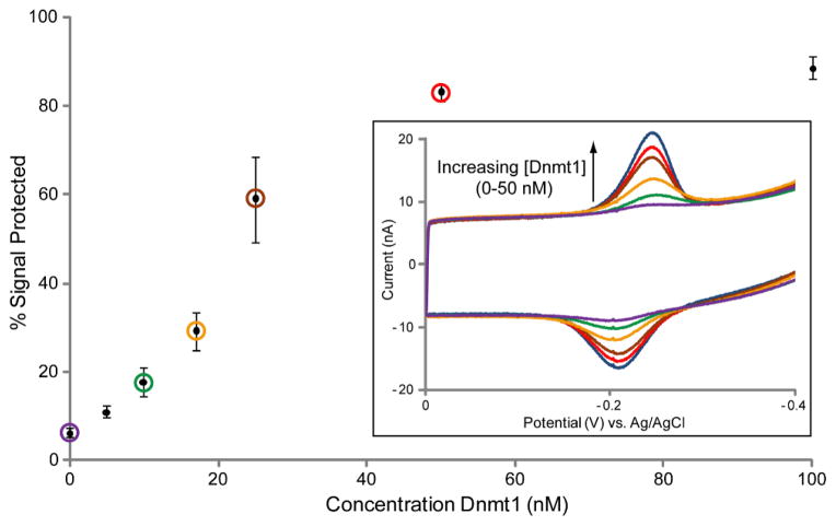
Figure 6.
Concentration dependence of Dnmt1 Methyltransferase activity. Chips were modified in all quadrants with the hemimethylated BssHII 22-mer. DNA protection by various concentrations of Dnmt1 was evaluated side by side on the same chip, and CV scans from representative electrodes are shown overlaid (inset). Overlaid CV traces include the initial signal before BssHII treatment (blue), 50 nM Dnmt1 (red), 25 nM Dnmt1 (brown), 17 nM Dnmt1 (orange), 10 nM Dnmt1 (green), and untreated (purple). Quantification of the observed signal protection is displayed as a Dnmt1 activity curve where the circled points correspond to the concentrations represented by the overlaid CV traces. Error bars represent the standard deviation across 4–12 electrodes tested for each Dnmt1 concentration on 4 chips. All CV scans were performed in optimized scanning buffer (5 mM phosphate, 50 mM NaCl, 4 mM MgCl2, 4 mM spermidine, 50 μM EDTA, 10% glycerol, pH 7) with an Ag/AgCl reference electrode at a 100 mV/s scan rate.

