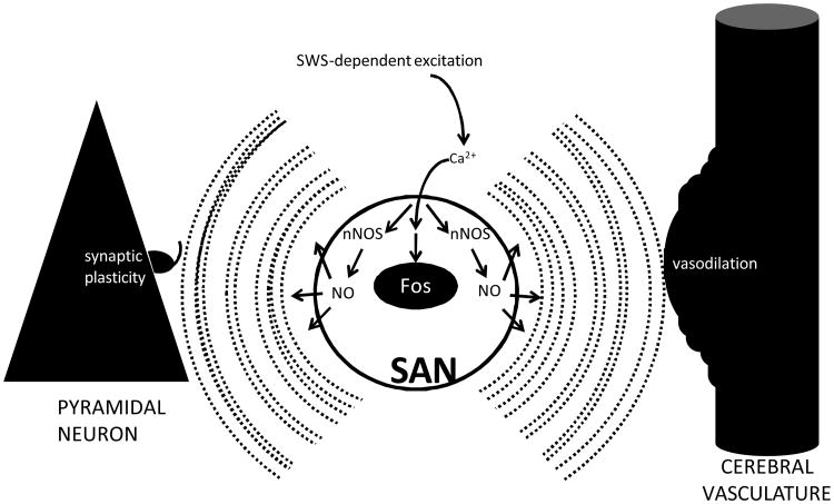
Figure 1.
Hypothetical model for the functions of SAN activation during SWS. At SWS onset, SANs are released from the tonic inhibition that they receive via monoaminergic and cholinergic inputs during wake. Excitation of SANs results in increased intracellular Ca2+ levels, which induces expression of Fos and activates nNOS. nNOS activation increases the concentration of NO within the cell. Being membrane permeant, NO disperses to nearby pyramidal cells. This increase in NO concentration is simultaneous with sleep-associated slow (approximately 1 Hz) oscillations in the electrical potential of pyramidal cells. The coincidence of 1 Hz oscillations and increased NO concentration induces long-term depression of synaptic inputs to the pyramidal cells (left). At the same time, NO triggers activity-dependent vasodilation of the cerebral microvasculature (right).

