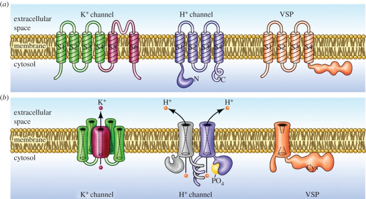
Figure 3.
Molecular architecture and orientation in the membrane of three classes of molecules that contain VSDs. (a) Illustrates the monomer of each, with its transmembrane segments numbered (S1, S2, etc.) starting from the intracellular N terminus. In each, the VSD includes the first four segments, S1–S4, in particular S4 which contains a series of cationic residues (Arg or Lys) that are thought to move outward upon membrane depolarization. The VSDs from these molecules are homologous. (b) Each molecule in its functional oligomeric state. The voltage-gated potassium (K+) channel is a tetramer, with four VSDs surrounding a single central pore, formed by the four S5–S6 segments, through which K+ crosses the membrane. The proton channel is a dimer in mammals and many other species, but each monomer has its own conduction pathway [8,9] and can function independently. Phosphorylation of Thr29 in the intracellular N terminus enhances the gating of HV in phagocytes and other cells [10,11]. The VSP is a monomer whose VSD senses membrane potential and accordingly regulates the activity of the attached intracellular phosphatase [12]. (Adapted from [13].)

