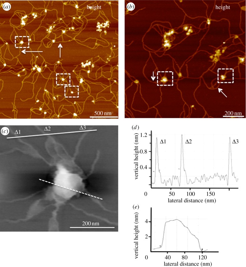
Figure 1.
PFM reveals glioblastoma exosomes with abundant nanofilamentous surface extensions. (a) Topographic image (z = 10 nm) of glioblastoma U87-derived exosomes showing round bulging vesicles (some shown in dashed boxes) surrounded by a network of nanofilaments. (b) The nanofilaments are shown at a higher resolution (z = 6 nm). Size of U87 exosomes measured from typical high-resolution PFM topographic image (c) is 89.3 nm in diameter and 4 nm in height (e; dashed white line). (d) Cross-section profiles show approximately 1.2 nm in height and 10 nm wide nanofilaments (marked Δ1–3, solid line). Fewer nanofilaments were seen in U251 exosomes, but not seen in NHA-derived exosomes (see the electronic supplementary material, figure S1). The results were confirmed by imaging samples obtained from two independent and commonly used isolations, with and without sucrose gradient purification. Size distributions show an average vesicle size of 89 ± 3.2 nm, 80.8 ± 2.2 nm and 70.9 ± 2.2 nm for U87, U251 and NHA, respectively (n ∼ 100 individual exosomes).

