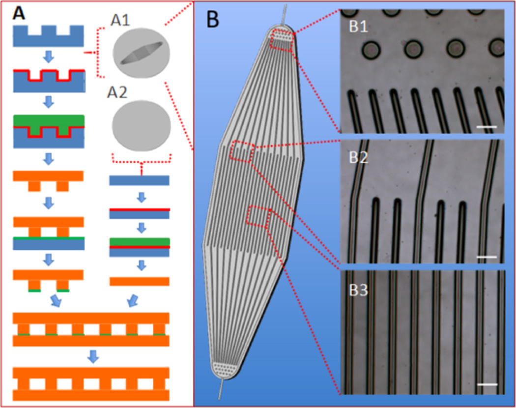
Figure 1.
Fabrication of PGS scaffolds from silicon wafers. (A) Assembly of a microfluidic device (µFD) base and thin membrane; (B) simplified schematic of the micropatterned wafer. (Insets A1 and A2) patterned and unpatterned wafers. (Insets B1–B3) light micrographs of µFD base showing transition from inlet posts to inlet channels (B1), transition from inlet region to central channels (B2), and central channels (B3). Silicon appears blue; maltose appears red; cured PGS appears orange, and PGS pre-polymer appears green. Scale bars: 100 µm.

