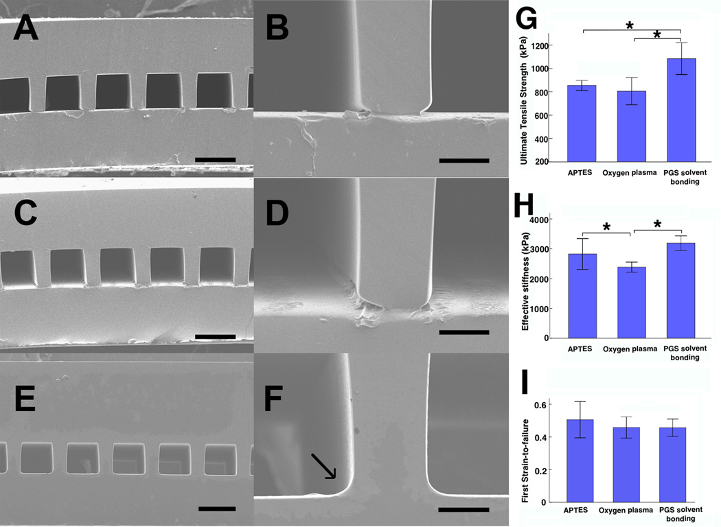
Figure 2.
Structural and mechanical properties of assembled scaffolds. (A–F) SEM images of scaffolds assembled by three methods (A, B) OP, (C, D) APTES and (E, F) PGS solvent bonding, which showed seamless binding (arrow) between the µFD base (above) and membrane (below). (G, H, I) Ultimate tensile stress (G), effective stiffness (H), and first strain-to-failure (I). Scale bars: (A, C, E) 100 µm, (B, D, F) 20 µm. Data are mean +/− SE. *Significant difference between groups.

