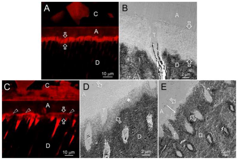
Figure 7.

A–E show adjacent confocal laser scanning microscopy (CLSM) and transmission electron microscopy images obtained from dentin bonds made with the self-etching dental adhesive, Adper Prompt-L-Pop. After bonding, the bonded tooth was sectioned vertically into 1 mm thick slabs and incubated in control media (Figs. A and B) or in biomimetic remineralization medium for 6 months. The specimen in Fig. A was immersed in 0.1% rhodamine B overnight. The red fluorescent tracer diffused into water-rich, resin-poor spaces within the hybrid layer (the area between the opposite arrows). In the absence of remineralizing reagents, no mineralization of the water-filled spaces occurred. The adjacent TEM shows no remineralization of the hybrid layer (area between opposing arrows). Fig. C is a CLSM image of the same resin-dentin bond that was allowed to remineralize for 6 months with biomimetic reagents. Note that the hybrid layer (area between opposing arrows) is less fluorescent and more grey indicating that much of the water was replaced by apatite mineral. This is better shown in the adjacent TEM images showing mineralization (asterisk region) of the bottom of the hybrid layer in 2 months and most of the hybrid layer in 6 months (from Kim et al. 2010 [166], with permission).
